The normal human heart contains 4 valves that regulate blood flow
into and out of the heart. The aortic and pulmonic valves are known as
the semilunar valves, whereas the tricuspid and mitral valves
are referred to as the atrioventricular valves. All the valves are
trileaflet, with the exception of the mitral valve, which has 2
leaflets. All 4 cardiac valves are surrounded by fibrous tissue forming
partial or complete valvular rings, or annuli. These annuli join the
fibrous skeleton of the heart to anchor and support the valvular
structures.
The aortic valve is located between the left ventricular outflow tract and the ascending aorta. It forms the centerpiece of the heart and closely approximates many other important cardiac structures (see the image below); specifically, the pulmonic valve anteriorly, mitral valve posterolaterally, and tricuspid valve posteromedially.[1]
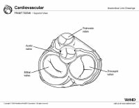 Heart valves, superior view. The
aortic valve functions to prevent the regurgitation of blood from the
aorta into the left ventricle during ventricular diastole and to allow
the appropriate flow of blood—the cardiac output
—from the left ventricle into the aorta during ventricular systole. The
aortic valve has 3 principle components: the annulus, cusps, and
commissures.
Heart valves, superior view. The
aortic valve functions to prevent the regurgitation of blood from the
aorta into the left ventricle during ventricular diastole and to allow
the appropriate flow of blood—the cardiac output
—from the left ventricle into the aorta during ventricular systole. The
aortic valve has 3 principle components: the annulus, cusps, and
commissures.
The dextrosuperior and sinistroinferior cushions fuse and, in doing so, form the truncal septum. The truncal septum undergoes a complex process of differentiation, eventually forming the right and left aortic valve cusps and 2 leaflets of the pulmonic valve. Of the 2 intercalated endocardial cushions, the right cushion eventually forms the posterior aortic valve cusp, whereas the left forms the anterior pulmonic valve leaflet. This occurs during the counterclockwise rotation and caudal shift of the conotruncus. During this time, the endocardial cushions also undergo dedifferentiation from a myosin-heavy chain to an alpha-smooth muscle actin phenotype, resulting in mature arterial valvular leaflets. The improper fusion or the incomplete dedifferentiation of the previously mentioned endocardial cushions is thought to be responsible for the formation of anatomically and structurally congenitally abnormal aortic valves.[2, 3, 4]
The following image is an overview of the transitions occurring in early heart development in amniotes.
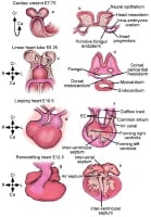 This
box provides an overview of the transitions occurring in early heart
development in amniotes (on the basis of the events in mouse
development). The whole embryo or isolated heart is shown on the left
(L). On the right (R), a representative section (transverse in panels b
and d; longitudinal in panels f and h) illustrates the main internal
features. As noted, staging in days of embryonic development (E) is
based on mouse development. The myocardium and its progenitors are
indicated in red. The cardiac progenitors are first recognizable as a
crescent-shaped epithelium (the cardiac crescent) at the cranial and
craniolateral parts of the embryo (panels a and b). The progenitor
population extends cranially and laterally almost to the junction
between the embryonic and extraembryonic regions of the embryo (red
arrow in panel b). Next, heart progenitors move ventrally to form the
linear heart tube (panels c and d). The linear heart tube undergoes a
complex progression termed cardiac looping, in which the tubular heart
adopts a spiral shape with its outer surface sweeping rightwards (panels
e and f). During looping, the inflow portion of the heart, including
the common atrium, is forced dorsally and cranially, so that it is now
above the developing ventricles. The internal relief of the heart at
this stage has become complex (panel f). Endocardial cushions (EC), the
precursors of the tricuspid and mitral valves (box 1), are forming in
the atrioventricular (AV) canal. Endocardial cushions also form in the
outflow tract, and these are the precursors of the aorticopulmonary
septum, which divides the outflow tract into the aorta and pulmonary
artery. These cushions also give rise to the aortic and pulmonary
valves. During the remodelling phase of heart development (panels g and
h), division of the heart chambers by septation is completed, and
distinct left (LV) and right ventricles (RV) and left (LA) and right
atria (RA) are evident. Ca = caudal (inferior); Cr = cranial (superior).
This
box provides an overview of the transitions occurring in early heart
development in amniotes (on the basis of the events in mouse
development). The whole embryo or isolated heart is shown on the left
(L). On the right (R), a representative section (transverse in panels b
and d; longitudinal in panels f and h) illustrates the main internal
features. As noted, staging in days of embryonic development (E) is
based on mouse development. The myocardium and its progenitors are
indicated in red. The cardiac progenitors are first recognizable as a
crescent-shaped epithelium (the cardiac crescent) at the cranial and
craniolateral parts of the embryo (panels a and b). The progenitor
population extends cranially and laterally almost to the junction
between the embryonic and extraembryonic regions of the embryo (red
arrow in panel b). Next, heart progenitors move ventrally to form the
linear heart tube (panels c and d). The linear heart tube undergoes a
complex progression termed cardiac looping, in which the tubular heart
adopts a spiral shape with its outer surface sweeping rightwards (panels
e and f). During looping, the inflow portion of the heart, including
the common atrium, is forced dorsally and cranially, so that it is now
above the developing ventricles. The internal relief of the heart at
this stage has become complex (panel f). Endocardial cushions (EC), the
precursors of the tricuspid and mitral valves (box 1), are forming in
the atrioventricular (AV) canal. Endocardial cushions also form in the
outflow tract, and these are the precursors of the aorticopulmonary
septum, which divides the outflow tract into the aorta and pulmonary
artery. These cushions also give rise to the aortic and pulmonary
valves. During the remodelling phase of heart development (panels g and
h), division of the heart chambers by septation is completed, and
distinct left (LV) and right ventricles (RV) and left (LA) and right
atria (RA) are evident. Ca = caudal (inferior); Cr = cranial (superior).
The aortic valve is located between the left ventricular outflow tract and the ascending aorta. It forms the centerpiece of the heart and closely approximates many other important cardiac structures (see the image below); specifically, the pulmonic valve anteriorly, mitral valve posterolaterally, and tricuspid valve posteromedially.[1]
Heart valves, superior view. The
aortic valve functions to prevent the regurgitation of blood from the
aorta into the left ventricle during ventricular diastole and to allow
the appropriate flow of blood—the cardiac output
—from the left ventricle into the aorta during ventricular systole. The
aortic valve has 3 principle components: the annulus, cusps, and
commissures. Embryology
Semilunar valve formation begins during the fourth week of gestation. At this time, opposing dextrosuperior and sinistroinferior endocardial cushions appear in the cephalad portion of the truncus arteriosus. Simultaneously, 2 additional intercalated endocardial cushions form, each located 90º from the aforementioned dextrosuperior and sinistroinferior endocardial cushions.The dextrosuperior and sinistroinferior cushions fuse and, in doing so, form the truncal septum. The truncal septum undergoes a complex process of differentiation, eventually forming the right and left aortic valve cusps and 2 leaflets of the pulmonic valve. Of the 2 intercalated endocardial cushions, the right cushion eventually forms the posterior aortic valve cusp, whereas the left forms the anterior pulmonic valve leaflet. This occurs during the counterclockwise rotation and caudal shift of the conotruncus. During this time, the endocardial cushions also undergo dedifferentiation from a myosin-heavy chain to an alpha-smooth muscle actin phenotype, resulting in mature arterial valvular leaflets. The improper fusion or the incomplete dedifferentiation of the previously mentioned endocardial cushions is thought to be responsible for the formation of anatomically and structurally congenitally abnormal aortic valves.[2, 3, 4]
The following image is an overview of the transitions occurring in early heart development in amniotes.
This
box provides an overview of the transitions occurring in early heart
development in amniotes (on the basis of the events in mouse
development). The whole embryo or isolated heart is shown on the left
(L). On the right (R), a representative section (transverse in panels b
and d; longitudinal in panels f and h) illustrates the main internal
features. As noted, staging in days of embryonic development (E) is
based on mouse development. The myocardium and its progenitors are
indicated in red. The cardiac progenitors are first recognizable as a
crescent-shaped epithelium (the cardiac crescent) at the cranial and
craniolateral parts of the embryo (panels a and b). The progenitor
population extends cranially and laterally almost to the junction
between the embryonic and extraembryonic regions of the embryo (red
arrow in panel b). Next, heart progenitors move ventrally to form the
linear heart tube (panels c and d). The linear heart tube undergoes a
complex progression termed cardiac looping, in which the tubular heart
adopts a spiral shape with its outer surface sweeping rightwards (panels
e and f). During looping, the inflow portion of the heart, including
the common atrium, is forced dorsally and cranially, so that it is now
above the developing ventricles. The internal relief of the heart at
this stage has become complex (panel f). Endocardial cushions (EC), the
precursors of the tricuspid and mitral valves (box 1), are forming in
the atrioventricular (AV) canal. Endocardial cushions also form in the
outflow tract, and these are the precursors of the aorticopulmonary
septum, which divides the outflow tract into the aorta and pulmonary
artery. These cushions also give rise to the aortic and pulmonary
valves. During the remodelling phase of heart development (panels g and
h), division of the heart chambers by septation is completed, and
distinct left (LV) and right ventricles (RV) and left (LA) and right
atria (RA) are evident. Ca = caudal (inferior); Cr = cranial (superior).


0 التعليقات:
إرسال تعليق