The anal canal is the most terminal
part of the lower GI tract/large intestine, which lies between the anal
verge (anal orifice, anus) in the perineum below and the rectum above.
Confusion and controversy exist regarding the anatomy of the anorectal
region in anatomy and surgical texts. The description in this topic is
from below upwards, as that is how this region is usually examined in
clinical practice. Images depicting the anal canal can be seen below.[1, 2]
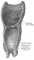 Coronal section of rectum and anal canal.
 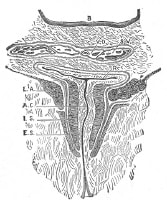 Coronal section through the anal canal. The
pigmented, keratinized perianal skin of the buttocks (around the anal
verge) has skin appendages (eg, hair, sweat glands, sebaceous glands);
compare this with the anal canal skin above the anal verge, which is
also pigmented and keratinized but does not have skin appendages.[3, 4]
Coronal section through the anal canal. The
pigmented, keratinized perianal skin of the buttocks (around the anal
verge) has skin appendages (eg, hair, sweat glands, sebaceous glands);
compare this with the anal canal skin above the anal verge, which is
also pigmented and keratinized but does not have skin appendages.[3, 4]
In anatomy texts, the rectum changes to the anal canal at the dentate line. For surgeons, however, the demarcation between the rectum above and the anal canal below is the anorectal ring. The following is the description of the surgical anal canal.
The anal canal is completely extraperitoneal. The length of the (surgical) anal canal is about 3-5 cm, with two thirds of this being above the dentate line and one third below the dentate line (anatomical anal canal).
The epithelium of the (anatomical) anal canal (between the anal verge below and the dentate line above) is variously described as anal mucosa or anal skin. The author feels that it should be called anal skin (anoderm), as it looks like (pigmented) skin, is sensitive like skin, and is keratinized (but does not have skin appendages).
The dentate line (also called the pectinate line) is the site of fusion of the proctodeum below and the postallantoic gut above. It is a wavy demarcation formed by the anal valves (transverse folds of mucosa) at the inferior-most ends of the anal columns. Anal glands open above the anal valves into the anal crypts. The dentate line is not seen on inspection in clinical practice, but under anesthesia the anal canal descends down, and the dentate line can be seen on slight retraction of the anal canal skin.
From a surgical perspective, the anal canal just above the dentate line for about 1-2 cm is called the transition zone. Beyond this transition zone, the (surgical) anal canal is lined with columnar epithelium. Anal columns (of Morgagni) are 5-10 longitudinal (vertical) mucosal folds in the upper part of the anal canal.
At the bottom of these columns are anal crypts, or sinuses, into which open the anal glands and anal papillae. Three of these columns (left lateral, right posterior, and right anterior, at 3, 7, and 11 o’clock position in supine position) are prominent; they are called anal cushions and contain branches and tributaries of superior rectal (hemorrhoidal) artery and vein. When prominent, veins in these cushions form the internal hemorrhoids.
The anorectal ring is situated about 5 cm from anus. At the anorectal angle, the rectum turns backwards to continue as the anal canal.
Levator ani and coccygeus muscles form the pelvic diaphragm. Lateral to the anal canal are the ischioanal fossae (1 on either side), below the pelvic diaphragm. The anterior relations of the anal canal are, in males, the seminal vesicles, prostate, and urethra, and, in females, the cervix and vagina. In front of (anterior to) the anal canal is the rectovesical fascia (of Denonvilliers), and behind (posterior) is the presacral endopelvic fascia (of Waldeyer), under which lie a rich presacral plexus of veins. Posterior to the anal canal lie the tip of the coccyx and lower sacrum.
Underneath the anal canal skin (below the dentate line) lies the external hemorrhoidal plexus of veins, which drains into systemic veins. Underneath the anal canal mucosa (above dentate line) lies the internal hemorrhoidal plexus of veins, which drains into the portal system of veins. The anorectum is, therefore, an important area of portosystemic venous connection (the other being the esophagogastric junction). Lymphatics from the anal canal drain into the superficial inguinal group of lymph nodes.
Coronal section of rectum and anal canal. Coronal section through the anal canal. The
pigmented, keratinized perianal skin of the buttocks (around the anal
verge) has skin appendages (eg, hair, sweat glands, sebaceous glands);
compare this with the anal canal skin above the anal verge, which is
also pigmented and keratinized but does not have skin appendages.[3, 4] In anatomy texts, the rectum changes to the anal canal at the dentate line. For surgeons, however, the demarcation between the rectum above and the anal canal below is the anorectal ring. The following is the description of the surgical anal canal.
The anal canal is completely extraperitoneal. The length of the (surgical) anal canal is about 3-5 cm, with two thirds of this being above the dentate line and one third below the dentate line (anatomical anal canal).
The epithelium of the (anatomical) anal canal (between the anal verge below and the dentate line above) is variously described as anal mucosa or anal skin. The author feels that it should be called anal skin (anoderm), as it looks like (pigmented) skin, is sensitive like skin, and is keratinized (but does not have skin appendages).
The dentate line (also called the pectinate line) is the site of fusion of the proctodeum below and the postallantoic gut above. It is a wavy demarcation formed by the anal valves (transverse folds of mucosa) at the inferior-most ends of the anal columns. Anal glands open above the anal valves into the anal crypts. The dentate line is not seen on inspection in clinical practice, but under anesthesia the anal canal descends down, and the dentate line can be seen on slight retraction of the anal canal skin.
From a surgical perspective, the anal canal just above the dentate line for about 1-2 cm is called the transition zone. Beyond this transition zone, the (surgical) anal canal is lined with columnar epithelium. Anal columns (of Morgagni) are 5-10 longitudinal (vertical) mucosal folds in the upper part of the anal canal.
At the bottom of these columns are anal crypts, or sinuses, into which open the anal glands and anal papillae. Three of these columns (left lateral, right posterior, and right anterior, at 3, 7, and 11 o’clock position in supine position) are prominent; they are called anal cushions and contain branches and tributaries of superior rectal (hemorrhoidal) artery and vein. When prominent, veins in these cushions form the internal hemorrhoids.
The anorectal ring is situated about 5 cm from anus. At the anorectal angle, the rectum turns backwards to continue as the anal canal.
Levator ani and coccygeus muscles form the pelvic diaphragm. Lateral to the anal canal are the ischioanal fossae (1 on either side), below the pelvic diaphragm. The anterior relations of the anal canal are, in males, the seminal vesicles, prostate, and urethra, and, in females, the cervix and vagina. In front of (anterior to) the anal canal is the rectovesical fascia (of Denonvilliers), and behind (posterior) is the presacral endopelvic fascia (of Waldeyer), under which lie a rich presacral plexus of veins. Posterior to the anal canal lie the tip of the coccyx and lower sacrum.
Blood supply and lymphatics
The anal canal above the dentate line is supplied by the terminal branches of the superior rectal (hemorrhoidal) artery, which is the terminal branch of the inferior mesenteric artery. The middle rectal artery (a branch of the internal iliac artery) and the inferior rectal artery (a branch of the internal pudendal artery) supply the lower anal canal.Underneath the anal canal skin (below the dentate line) lies the external hemorrhoidal plexus of veins, which drains into systemic veins. Underneath the anal canal mucosa (above dentate line) lies the internal hemorrhoidal plexus of veins, which drains into the portal system of veins. The anorectum is, therefore, an important area of portosystemic venous connection (the other being the esophagogastric junction). Lymphatics from the anal canal drain into the superficial inguinal group of lymph nodes.


0 التعليقات:
إرسال تعليق