Comprehensive knowledge of anatomy forms the basis for understanding
and treatment of neurological disease. Intracranial arteries are
involved in many neurologic disorders. Knowledge of arterial anatomy,
variants, and areas involved in disease is essential to define the
location of neurovascular lesions, delineate the extent and involvement
of branching perforators, and assess the effects on downstream
perfusion.
Arterial anatomy adds to the complexity of neurologic localization, providing a unique classification of neurovascular disorders. Arterial anatomy is also intertwined with pathophysiology, as vessel morphology influences hemodynamic variables. Only marginal advances regarding pathology of these arterial segments has been made since autopsy series performed hundreds of years ago.
Angiography and numerous noninvasive imaging methods have been developed to image intracranial arterial anatomy,[1, 2] yet these modern vascular imaging techniques including transcranial Doppler (TCD) ultrasonography, computed tomographic angiography (CTA), and magnetic resonance angiography (MRA) are not as accurate as the gold standard of conventional or digital subtraction angiography (DSA) (see the image below).
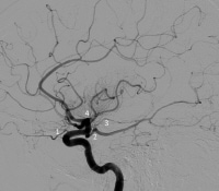 Lateral
projection of a left common carotid artery injection that displays the
order of branching in the intracranial carotid, including 1: ophthalmic,
2: posterior communicating, 3: anterior choroidal, and 4: anterior
cerebral arteries.
Lateral
projection of a left common carotid artery injection that displays the
order of branching in the intracranial carotid, including 1: ophthalmic,
2: posterior communicating, 3: anterior choroidal, and 4: anterior
cerebral arteries.
Arterial anatomy adds to the complexity of neurologic localization, providing a unique classification of neurovascular disorders. Arterial anatomy is also intertwined with pathophysiology, as vessel morphology influences hemodynamic variables. Only marginal advances regarding pathology of these arterial segments has been made since autopsy series performed hundreds of years ago.
Angiography and numerous noninvasive imaging methods have been developed to image intracranial arterial anatomy,[1, 2] yet these modern vascular imaging techniques including transcranial Doppler (TCD) ultrasonography, computed tomographic angiography (CTA), and magnetic resonance angiography (MRA) are not as accurate as the gold standard of conventional or digital subtraction angiography (DSA) (see the image below).
Lateral
projection of a left common carotid artery injection that displays the
order of branching in the intracranial carotid, including 1: ophthalmic,
2: posterior communicating, 3: anterior choroidal, and 4: anterior
cerebral arteries. 


0 التعليقات:
إرسال تعليق